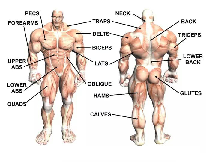
Lower Back Muscles Names / Muscles Of The Lumbar Spine Of The Trunk - The back anatomy includes the latissimus dorsi, trapezius, erector spinae, rhomboid, and the teres major.
Lower Back Muscles Names / Muscles Of The Lumbar Spine Of The Trunk - The back anatomy includes the latissimus dorsi, trapezius, erector spinae, rhomboid, and the teres major.. The teres major muscles work with the rotator cuff muscles to stabilize the shoulder joint and works with the latissimus dorsi muscles to pull the humerus back. On this page, you'll learn about each of these muscles, their locations and functional anatomy. The superficial group, the deep group, and the intermediate group. This muscle is the largest flexor of the foot. These muscles include the large paired muscles in the lower back, called erector spinae, which help hold up the spine, and gluteal muscles.
The gluteus maximus is one of the most important muscles in the body, and keeping it strong can help support the lower back. If you'd like to support us and get something great in return, check out our osce it is divided into three distinct segments, named the superior (s), middle (m) and inferior (i) lower brainstem and upper cervical cord lesions can interfere with the function of cranial nerve xi. Attached to the spine by soft tissues call tendons, these muscles control back motions, support the spine and. See back muscles and low back pain. The flexor muscles are attached to the front of the spine and enable flexing, bending forward, lifting, and arching the lower back.
Transversospinales Physiopedia from www.physio-pedia.com The muscles of the lower back help stabilize, rotate, flex, and extend the spinal column, which is a bony tower of 24 vertebrae that gives the body structure and houses the spinal cord. Muscle strains and sprains are common in the lower back, because it supports the weight of the upper body and is involved in moving, twisting and bending. The part of the nerve that emerges out of the spine is called the nerve root. All the extrinsic back muscles are innervated by the ventral (anterior) rami of the cervical spinal nerves , except for the trapezius muscle which receives its supply from the accessory nerve (cn xi) . Muscle anatomy in shoulder 12 photos of the muscle anatomy in shoulder muscle anatomy neck and shoulder, muscle anatomy of shoulder, muscle anatomy of shoulder joint, muscle anatomy shoulder back, muscle anatomy shoulder upper arm, human muscles, muscle anatomy neck and shoulder, muscle anatomy of shoulder, muscle. These muscles provide posture and stability to the body by holding the vertebral column erect and adjusting the position of the body to maintain balance. Its name means belly of the leg,and its common name is the calf muscle. This traditional yoga pose works your gluteus maximus, hamstrings, and spinal extensors.
The vertebral column of the lower back includes the five lumbar vertebrae, the sacrum, and the coccyx.
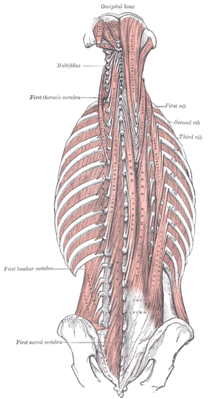
Anterior rami of lower thoracic nerves (t9 to t12). Five pairs of lumbar spinal nerves labeled l1 to l5 branch off your spinal cord and exit through small holes between the vertebrae. This muscle is the largest flexor of the foot. These muscles provide posture and stability to the body by holding the vertebral column erect and adjusting the position of the body to maintain balance. The superficial group, the deep group, and the intermediate group. These muscles include the large paired muscles in the lower back, called erector spinae, which help hold up the spine, and gluteal muscles. Its relaxing effect on your. Three main muscle groups are located in the lower back: Muscles of the lumbar spine. The back anatomy includes the latissimus dorsi, trapezius, erector spinae, rhomboid, and the teres major. The muscles of the lower back, including the erector spinae and quadratus lumborum muscles, contract to extend and laterally bend the vertebral column. This traditional yoga pose works your gluteus maximus, hamstrings, and spinal extensors. The deep or intrinsic muscles of the back.
The superficial group, the deep group, and the intermediate group. Lower border of ribs ix to xii just lateral to their angles. Its name means belly of the leg,and its common name is the calf muscle. Out of these, the cookies that are categorized as necessary are stored on your browser as they are essential for the working of basic functionalities of the website. Lumbar muscle strain is caused when muscle fibers are abnormally stretched or torn.
Hamstring Muscles And Your Back Pain from www.verywellhealth.com The vertebral column of the lower back includes the five lumbar vertebrae, the sacrum, and the coccyx. Nerves in your lower back. The part of the nerve that emerges out of the spine is called the nerve root. Lumbar muscle strain is caused when muscle fibers are abnormally stretched or torn. Lie on the ground and bend the knees, placing the. In the meanwhile, your hip flexors, quadriceps and lumbar muscles remain tight to keep you in an upright position. The pelvic floor muscles also help increase this pressure, which provides stability to the spine and trunk. Lumbar (lower back) muscle strains and sprains are the most common causes of low back pain.
The muscles of the lower back, including the erector spinae and quadratus lumborum muscles, contract to extend and laterally bend the vertebral column.
(2017, elsevier) should be consulted. All the extrinsic back muscles are innervated by the ventral (anterior) rami of the cervical spinal nerves , except for the trapezius muscle which receives its supply from the accessory nerve (cn xi) . Its relaxing effect on your. These muscles provide posture and stability to the body by holding the vertebral column erect and adjusting the position of the body to maintain balance. The rhomboids (major and minor) originate on the spinal column and attach to the middle (medial) surface of the scapula. Nerves in your lower back. The muscles of the lower back, including the erector spinae and quadratus lumborum muscles, contract to extend and laterally bend the vertebral column. Three main muscle groups are located in the lower back: Intermediate extrinsic muscles of the back: This traditional yoga pose works your gluteus maximus, hamstrings, and spinal extensors. Related posts of muscles of the lower back and buttocks diagram muscle anatomy drawing. Lumbar (lower back) muscle strains and sprains are the most common causes of low back pain. The muscles that move the upper legs (thigh) there are many muscles that move the large bone of the thigh.
The superficial group, the deep group, and the intermediate group. This portion also attaches to the back part of the transverse processes of. The quick answer to this question is the muscles of the lower back are the multifidus, longissimus, spinalis, and quadratus lumborum. Its name means belly of the leg,and its common name is the calf muscle. The quadratus lumborum muscles (orange, in the image above) are found in the lower back (also called the lumbar area).
Increasephysique from increasephysique.neocities.org Depresses ribs ix to xii and may prevent lower ribs from being elevated when the diaphragm contracts. Bones of the pelvis and lower back the bones of the pelvis and lower back work together to support the body's weight, anchor the abdominal and hip muscles, and protect the delicate vital organs of the vertebral and abdominopelvic cavities. The superficial group, the deep group, and the intermediate group. There are three different muscle groups found in the back: The thoracic portion also attaches to ribs, but these are the top part of the upper six ribs. Attached to the spine by soft tissues call tendons, these muscles control back motions, support the spine and. This blog post article is an overview of the muscles of the lumbar spine of the trunk. The flexor muscles are attached to the front of the spine and enable flexing, bending forward, lifting, and arching the lower back.
It is composed of trapezius, latissimus dorsi, rhomboid major, rhomboid minor and levator scapulae.
The gastrocnemius runs down the back of the lower leg, from the end of the femur to the heel bone, or calcaneus. All the extrinsic back muscles are innervated by the ventral (anterior) rami of the cervical spinal nerves , except for the trapezius muscle which receives its supply from the accessory nerve (cn xi) . It is innervated by anterior rami of spinal nerves, reflecting its embryological origin outside the back. Lumbar (lower back) muscle strains and sprains are the most common causes of low back pain. The lumbar portion of the iliocostalis muscle travels upward from the lower area of the pelvis and sacrum to attach onto the lower border of the bottom six or seven ribs, by means of tendons that branch off from the main line.; The quadratus lumborum muscles (orange, in the image above) are found in the lower back (also called the lumbar area). Three main muscle groups are located in the lower back: Its relaxing effect on your. The rhomboids (major and minor) originate on the spinal column and attach to the middle (medial) surface of the scapula. They help to bend the back to one side or the other. It is composed of trapezius, latissimus dorsi, rhomboid major, rhomboid minor and levator scapulae. Deep group of back muscles. Muscle strains and sprains are common in the lower back, because it supports the weight of the upper body and is involved in moving, twisting and bending.
These muscles provide posture and stability to the body by holding the vertebral column erect and adjusting the position of the body to maintain balance lower back muscles. Nerves in your lower back.

:max_bytes(150000):strip_icc()/GettyImages-87308179-56a05f563df78cafdaa14cd4.jpg)


0 Komentar